Staff
Prof. Dr. Benedikt Kost
Contact
Prof. Dr. Sabine Müller
Contact
PD Dr. Michael Lebert
Privatdozent
Contact
Dr.habil. Maria Ntefidou
Research associates
Contact
Dr. Peter Richter
Research associates
Contact
Joline Blaß
Research associates
Contact
Carolin Fritz
Research associates
Contact
Johanna Knab
Research associates
Contact
Choy Kriechbaum
Research associates
Contact
Dr. Sylwia Schulmeister
AG Kost
+49 9131 85 – 28219
sylwia.schulmeister [æt] fau.de
Jennifer Schuster
AG Müller
+49 9131 85 – 28224
jennifer.t.schuster [æt] fau.de
Martin Schuster
AG Kost
+49 9131 85 – 28230
martin.t.schuster [æt] fau.de
Sandra Tauber
AG Kost/AG Müller
+49 9131 85 – 28218 o. 28224
sandra.tauber [æt] fau.de
Hildegard Voll
AG Kost
+49 9131 85 – 28218
hildegard.voll [æt] fau.de
Norbert Meißner
Technischer Sekretär
+49 9131 85 – 28230 o. 28236
norbert.meissner [æt] fau.de
Prof. Dr. Georg Kreimer
georg.kreimer [æt] fau.de
Research
Photoorientation of Flagellate Green Algae
Our research is focused on the analysis of the eyespot apparatus, the „eye“, of flagellate green algae. Using a combination of biochemical, cell and molecular biological techniques we try to functional characterize this primordial visual system. Due to their elaborate structures and the presence of retinal-based photoreceptors in some lineages, algal eyespots are thought to play an important role in the evolution of photoreception. The eyespot apparatus of green algae utilizes specialized microbial-type rhodopsins as photoreceptors, which act as directly light-gated ion channels. After establishment of an isolation method for the eyespot apparatus in its entire complexity and its core and phosphoproteome (Schmidt et al. 2006, Plant Cell 18: 1908-1930; Wagner et al. 2008, Plant Physiol. 146: 772-788), we are now interested in the signal transduction cascades leading to the photoresponses of these algae and the mechanisms underlying the tight interaction of the different subcellular compartments in this region of the cell. Our current focus lies on the functional characterisation of novel Ca2+‑binding proteins and the analyses of protein-protein interactions and protein complexes. Another topic is focused on the funtional analysis of the photoreceptors channelrhodopsin 1 and phototropin. For this we are employing different experimental approaches like e.g. RNAi, phosphorylation analysis, immunoprecipitation and different 2D-electrophoretic approaches in combination with mass spectrometry.
Our main model organism is the green alga Chlamydomonas reinhardtii. Some advantages of this alga, except those already pointed out by Mike Adams („Chlamydomonas is pretty, non-pathogenic, doesn´t stink, doesn´t contaminate other cultures and no one cares if you kill it.”), are summarized here.
Some of the advantages of Chlamydomonas as a model system are listed below:
Although eukaryotic, standard microbiological techniques can be applied.
The cells can be grown either autothrophic, heterotrophic or mixothrophic in well defined liquid media or on agar. Culturing is easy and large amounts of cells can be grown under axenic conditions, e.g. we can grow up to 100 L of cultures per week.
Synchronized growth is possible.
C. reinhardtii includes asexual and sexual reproductions in its life cycle. The sexual reproduction can easily be induced by nitrogen starvation and blue light.
C. reinhardtii is genetically well characterized. All three genomes (nulceus, chloroplast, mitochondrium) are sequenced and can be transformed by simple techniques.
Diverse, large mutant collections are available. Mutants, BAC and cDNA libraries can be obtained via the Chlamydomonas Center.
The cells are motile and their different behavioural responses can be analyzed easily.
Many of ist proteins have higher homology to mamalian proteins than to those of higher plants. Hence Chlamydomonas is also a model for some human diseases, especially those related to flagella dysfunction.
The ultrastructure of its eyespot apparatus is depicted briefly in Fig. 1A. It is a singular structure of ~ 1 µm2 and can easily be detected in the light microscope due to the massive accumulation of carotenoids. Detailed ultrastructural and biophysical analyses have shown that the functional eyespot apparatus involves local specialisations of different subcellular compartments (plasma membrane, cytosol, and chloroplast).
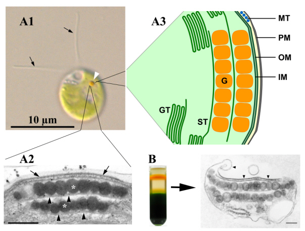
Fig. 1: The eyespot apparatus of Chlamydomonas reinhardtii in the light microscope (A1, white arrow-head), in the transmission electron microscope (A2) and as a schematic drawing (A3). Structural intact eyespot apparatuses can be isolated by sucrose density gradient centrifugation (B).
The ultrastructure of the functional eyespot apparatus is complex and involves local specializations of membranes from different compartments. In Chlamydomonas reinhardtii, the eyespot apparatus is usually composed of two highly ordered layers of carotenoid-rich lipid globules inside the chloroplast (Figure 1A ). The globules exhibit a remarkably constant diameter of 80 to 130 nm and are subtended by a thylakoid membrane. Additionally, the outermost globule layer is attached to specialized areas of the two chloroplast envelope membranes and the adjacent plasma membrane. This whole complex can be isolated as a structural unit (Fig. 1B). The plasma membrane and the outer chloroplast envelope membrane above the eyespot globules are extremely rich in intramembrane particles. The photoreceptors identified so far are generally believed to be located in this plasma membrane patch. Light-dependent oriented movement responses (phototaxis) require the cell to determine the direction of incident light. Chlamydomonas most likely accomplishes this by monitoring the modulation of the light intensity reaching its photoreceptors in the eyespot apparatus as the cell rolls around its longitudinal cell axis during helical forward swimming. Increasing evidence points to additional important roles of the eyespot apparatus beside those in light perception.
A more detailed description of the structure and function of this algal “eye” can be found in the following review (Kreimer 2009, Curr. Genet. 55, 19-43).
Publications
Authored Books
- Kreimer, G., Wakabayashi, K.I., Hegemann, P., & Dieckmann, C. (2023). The eyespot and behavioral light responses. Elsevier.
BibTeX: Download
Journal Articles
- Wolfram, M., Greif, A., Baidukova, O., Voll, H., Tauber, S., Lindacher, J.,... Kreimer, G. (2024). Insights into degradation and targeting of the photoreceptor channelrhodopsin-1. Plant Cell and Environment. https://doi.org/10.1111/pce.15017
BibTeX: Download - Wolfram, M., Greif, A., Sizova, I., Baidukova, O., Hegemann, P., & Kreimer, G. (2023). Multifactorial in vivo regulation of the photoreceptor channelrhodopsin-1 abundance. Plant Cell and Environment. https://doi.org/10.1111/pce.14657
BibTeX: Download - Böhm, M., Boness, D., Fantisch, E., Erhard, H., Frauenholz, J., Kowalzyk, Z.,... Kreimer, G. (2019). Channelrhodopsin-1 phosphorylation changes with phototactic behavior and responds to physiological stimuli in Chlamydomonas. The Plant Cell. https://doi.org/10.1105/tpc.18.00936
BibTeX: Download - Greiner, A., Kelterborn, S., Evers, H., Kreimer, G., Sizova, I., & Hegemann, P. (2017). Targeting of Photoreceptor Genes in Chlamydomonas reinhardtii via Zinc-finger Nucleases and CRISPR/Cas9. The Plant Cell, 29(10), 2498-2518. https://doi.org/10.1105/tpc.17.00659
BibTeX: Download - Trippens, J., Reißenweber, T., & Kreimer, G. (2017). The chloroplast Calcium Sensor protein CAS affects phototactic behaviour in Chlamydomonas reinhardtii (Chlorophyceae) at low light intensities. Phycologia, 56(3), 261-270. https://doi.org/10.2216/16-67.1
BibTeX: Download - Eitzinger, N., Wagner, V., Weisheit, W., Geimer, S., Boness, D., Kreimer, G., & Mittag, M. (2015). Proteomic Analysis of a Fraction with Intact Eyespots of Chlamydomonas reinhardtii and Assignment of Protein Methylation. Frontiers in Plant Science, 6. https://doi.org/10.3389/fpls.2015.01085
BibTeX: Download - Schulze, T., Schreiber, S., Iliev, D., Boesger, J., Trippens, J., Kreimer, G., & Mittag, M. (2013). The Heme-Binding Protein SOUL3 of Chlamydomonas reinhardtii Influences Size and Position of the Eyespot. Molecular Plant, 6(3), 931-944. https://doi.org/10.1093/mp/sss137
BibTeX: Download - Trippens, J., Greiner, A., Schellwat, J., Neukam, M., Rottmann, T., Lu, Y.,... Kreimer, G. (2012). Phototropin Influence on Eyespot Development and Regulation of Phototactic Behavior in Chlamydomonas reinhardtii. The Plant Cell, 24(11), 4687-4702. https://doi.org/10.1105/tpc.112.103523
BibTeX: Download - Rolland, N., Atteia, A., Decottignies, P., Garin, J., Hippler, M., Kreimer, G.,... Wagner, V. (2009). Chlamydomonas proteomics. Current Opinion in Microbiology, 12(3), 285-291. https://doi.org/10.1016/j.mib.2009.04.001
BibTeX: Download - Kreimer, G. (2009). The green algal eyespot apparatus: a primordial visual system and more? Current Genetics, 55(1), 19-43. https://doi.org/10.1007/s00294-008-0224-8
BibTeX: Download - Wagner, V., Ullmann, K., Mollwo, A., Kaminski, M., Mittag, M., & Kreimer, G. (2008). The phosphoproteome of a Chlamydomonas reinhardtii eyespot fraction includes key proteins of the light signaling pathway. Plant Physiology, 146(2), 772-788. https://doi.org/10.1104/pp.107.109645
BibTeX: Download - Wagner, V., Kreimer, G., & Mittag, M. (2008). The power of functional proteomics: Components of the green algal eyespot and its light signaling pathway(s). Plant Signaling & Behavior, 3(7), 433-435. https://doi.org/10.4161/psb.3.7.5685
BibTeX: Download - Kreimer, G. (2007). Evidence for a specialized localization of the chloroplast ATP-synthase subunits alpha, beta, and gamma in the eyespot apparatus of Chlamydomonas reinhardtii (Chlorophyceae). Journal of Phycology, 43(2), 284-294. https://doi.org/10.1111/j.1529-8817.2007.00331.x
BibTeX: Download - Kreimer, G. (2006). Proteomic analysis of the eyespot of Chlamydomonas reinhardtii provides novel insights into its components and tactic movements. The Plant Cell, 18(8), 1908-1930. https://doi.org/10.1105/tpc.106.041749
BibTeX: Download - Kreimer, G. (2006). Toward a protein map of the green algal eyespot: analysis of eyespot globule-associated proteins. Phycologia, 45(2), 199-212.
BibTeX: Download - Renninger, S., Backendorf, E., & Kreimer, G. (2001). Subfractionation of eyespot apparatuses from the green alga |Spermatozopsis similis|: Isolation and characterization of eyespot globules. Planta, 213, 51-63. https://dx.doi.org/10.1007/s004250000473
BibTeX: Download - Hill, K., Hemmler, R., Kovermann, P., Calenberg, M., Kreimer, G., & Wagner, R. (2000). A Ca2+- and voltage-modulated flagellar ion channel is a component of the mechanoshock response in the unicellular green alga Spermatozopsis similis. Biochimica Et Biophysica Acta-Biomembranes, 1466(1-2), 187-204. https://dx.doi.org/10.1016/S0005-2736(00)00200-5
BibTeX: Download - Dole, V., Jakubzik, C.R., Brünjes, B., & Kreimer, G. (2000). A cDNA from the green alga Spermatozopsis similis encodes a protein with homology to the newly discovered Roadblock/LC7 family of dynein-associated proteins. Gene Structure and Expression, 1490(1-2), 125-130. https://dx.doi.org/10.1016/S0167-4781(99)00220-1
BibTeX: Download - Kreimer, G. (1999). Reflective properties of different eyespot types in dinoflagellates. Protist, 150, 311-323. https://dx.doi.org/10.1016/S1434-4610(99)70032-5
BibTeX: Download - Calenberg, M., Brohsonn, U., Zedlacher, M., & Kreimer, G. (1998). Light- and Ca2+-modulated heterotrimeric GTPases in the eyespot apparatus of a flagellate green alga. The Plant Cell, 10(1), 91-103. https://dx.doi.org/10.1105/tpc.10.1.91
BibTeX: Download - Santos, L.M.A., Melkonian, M., & Kreimer, G. (1996). A combined reflection confocal laser scanning, electron and fluorescence microscopy analysis of the eyespot in zoospores of Vischeria spp. (Eustigmatales, Eustigmatophyceae). Phycologia, 35(4), 299-307. https://dx.doi.org/10.2216/i0031-8884-35-4-299.1
BibTeX: Download - Kreimer, G. (1996). Light reception and signal modulation during photoorientation of flagellate green algae. Photochemistry and Photobiology, 63(1), 62-63.
BibTeX: Download - Schlicher, U., Linden, L., Calenberg, M., & Kreimer, G. (1995). G proteins and Ca2+-modulated protein kinases of a plasma membrane-enriched fraction and isolated eyespot apparatuses of Spermatozopsis similis (Chlorophyceae). European Journal of Phycology, 30(4), 319-330. https://dx.doi.org/10.1080/09670269500651111
BibTeX: Download - Linden, L., & Kreimer, G. (1995). Calcium modulates rapid protein phosphorylation/dephosphorylation in isolated eyespot apparatuses of the green alga Spermatozopsis similis. Planta, 197(2), 343-351. https://dx.doi.org/10.1007/BF00202656
BibTeX: Download - Kreimer, G. (1995). Flagellar Shock Responses in Green Algae. Botanica Acta : Berichte der Deutschen Botanischen Gesellschaft, 108(3), 169-171. https://dx.doi.org/10.1111/j.1438-8677.1995.tb00847.x
BibTeX: Download - Calenberg, M., Linden, L., & Kreimer, G. (1995). Ca2+ stimulated protein kinases and G-proteins are enriched in both, a plasma membrane fraction and isolated eyespot apparatuses of a green alga. European Journal of Cell Biology.
BibTeX: Download - Höning, S., Kreimer, G., Robenek, H., & Jockusch, B.M. (1994). Receptor-mediated endocytosis is sensitive to antibodies against the uncoating ATPase (hsc70). Journal of Cell Science, 107, 1185-1196.
BibTeX: Download - Grung, M., Kreimer, G., Calenberg, M., Melkonian, M., & Liaaen-Jensen, S. (1994). Carotenoids in the eyespot apparatus of the flagellate green alga Spermatozopsis similis: Adaptation to the retinal-based photoreceptor. Planta, 193(1), 38-43. https://dx.doi.org/10.1007/BF00191604
BibTeX: Download - Kreimer, G., & Witman, G.B. (1994). Novel touch-induced, Ca2+-dependent phobic response in a flagellate green alga. Cell Motility and the Cytoskeleton, 29(2), 97-109. https://dx.doi.org/10.1002/cm.970290202
BibTeX: Download - Hesse, T., Garbers, C., Brzobohaty, B., Kreimer, G., Söll, D., Melkonian, M.,... Palme, K. (1993). Two members of the ERabp gene family are expressed differentially in reproductive organs but to similar levels in the coleoptile of maize. Plant Molecular Biology, 23(1), 57-66. https://dx.doi.org/10.1007/BF00021419
BibTeX: Download - Kreimer, G., Overländer, C., Sineshchekov, O.A., Stolzis, H., Nultsch, W., & Melkonian, M. (1992). Functional analysis of the eyespot in Chlamydomonas reinhardtii mutant ey 627, mt−. Planta, 188(4), 513-521. https://dx.doi.org/10.1007/BF00197043
BibTeX: Download - Kreimer, G., Marner, F.-J., Brohsonn, U., & Melkonian, M. (1991). Identification of 11-cis and all-trans-retinal in the photoreceptive organelle of a flagellate green alga. Febs Letters, 293(1-2), 49-52. https://dx.doi.org/10.1016/0014-5793(91)81150-7
BibTeX: Download - Kreimer, G., Brohsonn, U., & Melkonian, M. (1991). Isolation and partial characterization of the photoreceptive organelle for phototaxis of a flagellate green alga. European Journal of Cell Biology, 55(2), 318-327.
BibTeX: Download - Höning, S., Jockusch, B.M., Kreimer, G., Veltel, D., Robenek, H., Engelhardt, W., & Frey, J. (1991). Endocytosis of human IgG: Fc receptor complexes by transfected BHK cells. European Journal of Cell Biology, 55(1), 48-59.
BibTeX: Download - Katsaros, C., Kreimer, G., & Melkonian, M. (1991). Localization of Tubulin and a Centrin-Homologue in Vegetative Cells and Developing Gametangia of Ectocarpus siliculosus (Dillw.) Lyngb. (Phaeophyceae, Ectocarpales) - A combined immunofluorescence and confocal laser scanning microscope study. Botanica Acta : Berichte der Deutschen Botanischen Gesellschaft, 104(2), 87-92. https://dx.doi.org/10.1111/j.1438-8677.1991.tb00201.x
BibTeX: Download - Kreimer, G., Kawai, H., Müller, D.G., & Melkonian, M. (1991). Reflective properties of the stigma in male gametes of Ectocarpus siliculosus (Phaeophyceae) studied by confocal laser scanning microscopy. Journal of Phycology, 27(2), 268-276. https://dx.doi.org/10.1111/j.0022-3646.1991.00268.x
BibTeX: Download - Kreimer, G., & Melkonian, M. (1990). Reflection confocal laser scanning microscopy of eyespots in flagellated green algae. European Journal of Cell Biology, 53(1), 101-111.
BibTeX: Download - Kreimer, G., Melkonian, M., Holtum, J.A.M., & Latzko, E. (1988). Stromal Free Calcium Concentration and Light-Mediated Activation of Chloroplast Fructose-1,6-Bisphosphatase. Plant Physiology, 86(2), 423-428. https://dx.doi.org/10.1104/pp.86.2.423
BibTeX: Download - Surek, B., Kreimer, G., Melkonian, M., & Latzko, E. (1987). Spinach ferredoxin is a calcium-binding protein. Planta, 171(4), 565-568. https://dx.doi.org/10.1007/BF00392307
BibTeX: Download - Kreimer, G., Surek, B., Woodrow, I.E., & Latzko, E. (1987). Calcium binding by spinach stromal proteins. Planta, 171(2), 259-265. https://dx.doi.org/10.1007/BF00391103
BibTeX: Download - Heimann, K., Kreimer, G., Melkonian, M., & Latzko, E. (1987). Light-Induced Ca2+ Influx into Spinach Protoplasts. Zeitschrift für Naturforschung Section C - A Journal of Biosciences, 42(3), 283-287.
BibTeX: Download - Kreimer, G., Melkonian, M., & Latzko, E. (1985). An electrogenic uniport mediates light-dependent Ca2+ influx into intact spinach chloroplasts. Febs Letters, 180(2), 253-258. https://dx.doi.org/10.1016/0014-5793(85)81081-4
BibTeX: Download - Kreimer, G., Melkonian, M., Holtum, J.A.M., & Latzko, E. (1985). Characterization of calcium fluxes across the envelope of intact spinach chloroplasts. Planta, 166(4), 515-523. https://dx.doi.org/10.1007/BF00391276
BibTeX: Download - Kreimer, G., & Latzko, E. (1983). Properties of l-ascorbate-using peroxidases from Pisum sativum L. Hoppe-Seyler's Zeitschrift für Physiologische Chemie, 364(9), 1165-1165.
BibTeX: Download
Book Contributions
- Kreimer, G., Wakabayashi, K.-i., Hegemann, P., & Dieckmann, C. (2023). The eyespot and behavioral light responses. In Dutcher S.K. (Eds.), The Chlamydomonas Sourcebook. (pp. 391-419). Elsevier Academic Press.
BibTeX: Download - Böhm, M., & Kreimer, G. (2020). Orient in the World with a Single Eye: The Green Algal Eyespot and Phototaxis. In Progress in Botany. (pp. 259-304). Cham: Springer.
BibTeX: Download - Kreimer, G. (2001). Light reception and signal modulation during photoorientation of flagellate green algae. In Donat-P. Häder and A.M. Breure (Eds.), Photomovement. (pp. 193-227). Amsterdam, London, New York, Oxford, Paris, Shannon, Tokyo: Elsevier.
BibTeX: Download - Kawai, H., & Kreimer, G. (2000). Sensory mechanisms: Light perception and taxis in algae. In Barry S. C. Leadbeater, John C. Green (Eds.), The Flagellates: Unity, Diversity and Evolution. (pp. 124-146). London and New York: Taylor & Francis.
BibTeX: Download - Kreimer, G. (1994). Cell Biology of Phototaxis in Flagellate Algae. In International Review of Cytology. (pp. 229-310). Academic Press Inc Ltd.
BibTeX: Download - Melkonian, M., Burchert, M., Kreimer, G., & Latzko, E. (1990). Binding and possible function of calcium in the chloroplast. In D.D. Randal and D.G. Blevins (Eds.), Current Topics in Plant Biochemistry and Physiology. (pp. 38-46). Interdisciplinary Plant Biochemistry and Physiology Program, University of Missouri-Columbia.
BibTeX: Download - Burchert, M., Surek, B., Kreimer, G., & Latzko, E. (1990). Calcium binding by chloroplast stroma proteins and functional implications. In Robert Thomas Leonard, Robert K. Hepler (Eds.), Calcium in plant growth and development: proceedings, 13th Annual Riverside Symposium in Plant Physiology. (pp. 17-25). American Society of Plant Physiologists.
BibTeX: Download - Latzko, E., Surek, B., & Kreimer, G. (1988). Calcium binding by stromal proteins and functional implications. In S. Vaklinova, V. Stanev and M. Dilova (Eds.), International Symposium on Plant Mineral Nutrition and Photosynthesis. (pp. 79-93).
BibTeX: Download - Kreimer, G., Surek, B., Heimann, K., Burchert, M., Lukow, L., Holtum, J.A.M.,... Latzko, E. (1987). Calcium metabolism in chloroplasts and protoplasts. In J. Biggins (Eds.), Proceedings of the 7th International Congress on Photosynthesis. (pp. 345-357). Dordrecht: Martinus Nijhoff Publishers.
BibTeX: Download - Latzko, E., Surek, B., & Kreimer, G. (1986). Ca2+ fluxes across the chloroplast envelope and Ca2+ binding by stromal proteins. In M. Gibbs (Eds.), Hungarian-USA Binational Symposium on Photosynthesis. (pp. 127-135).
BibTeX: Download

(Enlarge)
(Enlarge)

(Enlarge)
(Enlarge)

(Enlarge)

(Enlarge)
(Enlarge)
(Enlarge)

(Enlarge)

(Enlarge)
(Enlarge)

(Enlarge)